


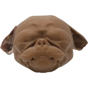
Description
Introducing Med Dimensions’ Canine Stifle, designed specifically for medical training and education in the field of veterinary medicine. This anatomically accurate model provides a comprehensive representation of the canine stifle joint, complete with major ligaments and tendons, to enhance learning and skill development.
Key Features:
- Realistic Anatomy: Our canine stifle bone model is meticulously crafted to mimic the intricate structures and proportions of the stifle joint found in canines. It accurately represents the bones, major ligaments, and tendons involved in the joint, providing an unparalleled training experience.
- Major Ligaments and Tendons: The model includes all the major ligaments and tendons associated with the canine stifle joint, such as the cranial cruciate ligament (CCL), caudal cruciate ligament (CrCL), medial collateral ligament (MCL), lateral collateral ligament (LCL), and the patellar tendon. These vital structures are clearly identifiable and can be studied in detail for a comprehensive understanding.
- Durable Construction: Made from high-quality materials, our canine stifle bone model is built to withstand rigorous training sessions. It offers longevity and reliability, ensuring that it can be used to cut and drill into bone without compromising its anatomical integrity.
- Interactive Learning: The model is designed to facilitate hands-on learning and interactive training. It allows students, veterinarians, and aspiring veterinary professionals to practice various procedures, such as ligament repairs, and surgical techniques, in a realistic and controlled environment.
- Educational Tool: Our canine stifle bone model is an invaluable educational tool that aids in the comprehension of the stifle joint’s complex anatomy and function. It serves as a visual aid during lectures, seminars, and workshops, enabling instructors to effectively teach and demonstrate crucial techniques and concepts.
- Versatile and Portable: The compact and lightweight design of the model makes it highly portable, allowing for easy transportation and convenient use in different learning environments. Whether in a classroom, veterinary clinic, or laboratory, this model adapts to various settings.
Enhance your veterinary training and prepare for real-life scenarios with our Canine Stifle Model. By providing a comprehensive representation of the stifle joint’s major ligaments and tendons, this model empowers veterinary professionals to develop their skills, deepen their knowledge, and ultimately deliver the highest standard of care to their canine patients.
MD-E-2000
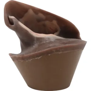
This product is a model of a left hind limb, derived from real CT scans to provide as close to real anatomy as possible.
The model includes CCL, LCL, MCL, Meniscus, Patellar Tendon, and Tendon of Ider constructs. It is a replica of a small/medium canine, roughly a 15-20kg dog, and is 8.5 inches long.
The ligaments are constructed of a blend of silicone that allows for proper constricted motion of the joint, as verified by leading veterinarians. The partial tibia and femur is 3D printed in a filament that is ideal for cutting and drilling practice. The silicone ligaments will hold suture, and are secured within the bone- not glued on- to prevent premature ripping of the ligaments from the insertion sites. The bone holds anchors, buttons, plates, and screws, providing tactile feedback to the user and ideal for demonstrations.
Reviews
There are no reviews yet.