Blog | Education | Technology
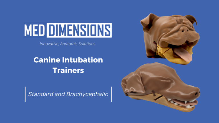
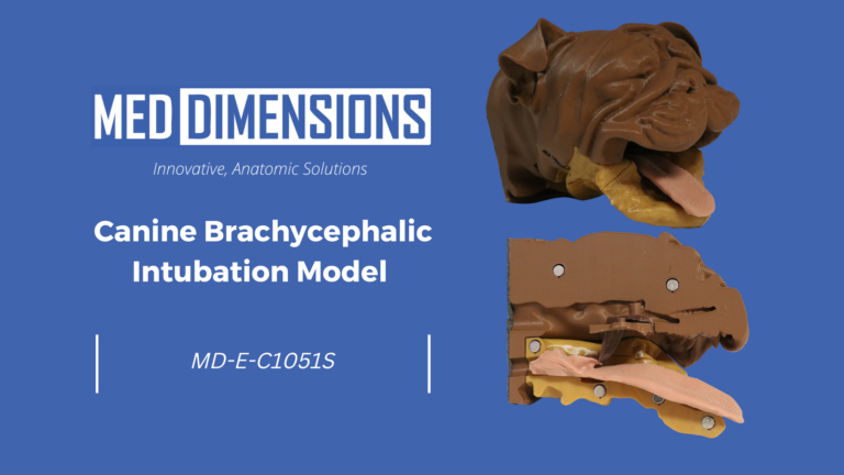
Canine Brachycephalic Intubation Trainers
Med Dimensions is excited to announce the release of our latest veterinary simulation model, the Canine Brachycephalic Intubation Trainer Bisected (MD-E-C1051S). This state-of-the-art model is designed to enhance the training and skills of veterinary professionals, offering a realistic and practical solution for canine intubation practice. Product Overview The MD-E-C1051S is a meticulously designed model of…