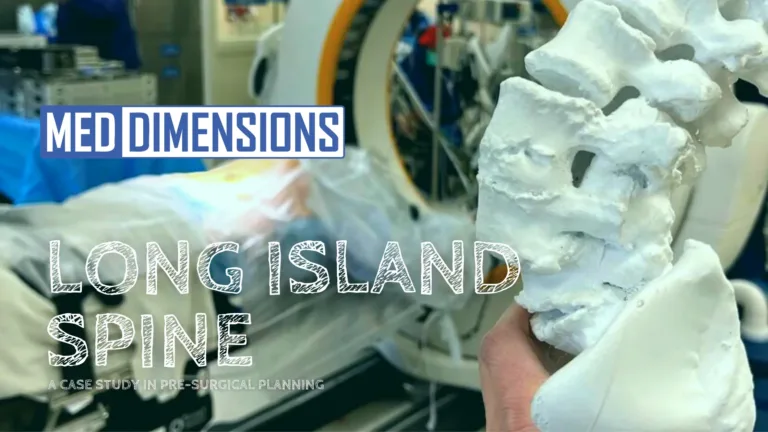
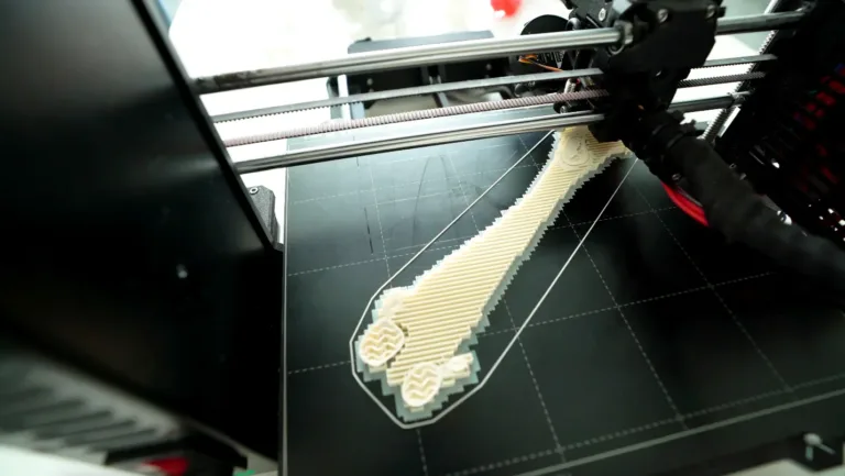
Disrupting Medical Education and Surgical Procedures with 3D Printing

Med Dimensions produces 3D models for the education, preparation, and assistance of surgical procedures for veterinary doctors, clinicians, and teachers.

From Chris Morgan at Matterhackers, Inc. – March 22, 2022

Located near Rochester, New York, Med Dimensions is a small startup created by two pet-loving engineers, Sean Bellefeuille and Will Byron, from Rochester Institute of Technology in 2019. Started as a campus club that was dedicated to 3D printing and helping others, Med Dimensions is a team that consists of professional designers, engineers, and business specialists that strive to find ways to improve the lives of surgeons, pets, and pet owners.

Focused primarily on veterinary medicine, the Med Dimensions team produces three-dimensional models for the education, preparation, and assistance in surgical procedures for veterinary doctors, clinicians, and teachers.

TPLO Guides and Trainers

The first focus of Med Dimensions, education, is geared toward creating models for the classroom to instruct a new generation of vets to be more prepared for common, and uncommon, surgical procedures. Vet students are sometimes deprived of hands-on training in many facets of a typical practice. Med Dimensions takes real anatomy and pathology and transforms it into a three-dimensional model for any type of procedure in any specialty, providing vital hands-on models for everything from bronchoscopies, intubations, suturing pads, dental trainers, and more.

Armed with more relevant, real-world models gives a new generation of vet students a leg up on previous classes. Using different materials in the fabrication of their educational models, Med Dimensions is able to replicate the look, feel, and performance of real anatomy. These models can be manipulated to test implant placement, practice techniques, and build confidence to treat animals safely and more effectively.

Educational Consult with Med Dimensions 3D Models

The second focus is preparation; Med Dimensions can create pre-operative models for specific cases pre-surgery so the veterinarian is able to see and feel what is occurring before surgery Because they use materials that feel and behave like real pathology, pre-operative models can be used for practice or for reference prior to surgery, saving time and money in the operating room. Director of Business Development, Michael Campbell explains, “From the beginning, Med Dimensions started this endeavor to help those involved in the surgery of animals get great outcomes.”

“Sometimes it’s simply not enough to see a two-dimensional scan and get all the information you need going into surgery. The best outcomes are served by three-dimensional, real-world scanned and 3D printed models that a surgeon can hold in their hand and not just have an educated guide, but an exact replica of an issue before making a single incision. That’s the kind of power we want to give to the teacher, the clinician, and the surgeon – we want them to be assured that they have the absolute best information about a procedure so they can affect the best outcome possible. We are also sensitive to the fact that having a veterinary practice is a business. Using our models, the clinicians are able to save time on many surgeries, as well as reduce the cost of those surgeries. They also enhance patient outcomes, making many more pet owners happy that their pets are feeling better post-op.”

Their third focus is, of course, performing surgeries. Not only do Med Dimensions create pre-operative models for general and specific surgeries, but they also work with veterinarians to fabricate patient-specific cutting guides and other custom surgical tools that can save time and money for patients and facilities alike. Shaving 20-30 minutes off a procedure, replicated over several procedures a year results in thousands of dollars in savings.

There are also technical advantages these tools can provide. A custom hip arthroplasty cutting guide can make it easier to replicate good cuts and measurements, allowing a surgeon to achieve better and more predictable outcomes in the operating room. In addition, a custom cutting guide can help steer a surgeon away from important structures in a certain area, specifically nerves, and arteries near a joint space.

Med Dimensions also works with the renowned surgeon, Dr. Johnny Uday, a leader in 3D printed guides and implants, to bring custom designs for surgical tools to life. With many years of experience in building custom implants in the human and veterinary fields, he speaks on the use of 3D modeling in the medical field, specializing in custom surgical applications.

From a business standpoint, the engineers and designers at Med Dimensions use their skills to help vet clinicians gain the best outcomes for their patients. As a ‘third arm’ in surgical preparation, Med Dimensions provides a cutting-edge service that up until recently, was almost impossible to do on an individual patient by patient basis.

Med Dimensions Pre-Op Model for Dr. Laurence Mermelstein at Long Island Spine Specialists (human)

Michael Campbell explains, “Again we look at scalability and also being extremely agile – previously to get a specific, custom model for a patient would have outpaced the cost of the surgery and the time turnaround was monumental – months on average. There simply were no manufacturing capabilities to quickly turn out an anatomically correct model at this low cost that we see now. The 3D printers, the software, and the materials – not to mention the engineers and designers that implement the process – none of those was within a reasonable cost in the past. Now with fast, in-house additive manufacturing of one-off models for specific patients, as well as using multiple farm printers to create stable educational models, all without the need to keep stock on the shelf or order massive quantities of materials to produce products that may never even be used, we’ve taken a very needed component of veterinary medicine, and soon human medicine, and made it affordable and sustainable.”

There are no giant warehouses we need to fill, we create models and custom models as needed and get them out the door. We don’t need shelves full of products for our doctors and clinicians to see more positive outcomes for their patients. That saves them time and money and it saves us time and money – it’s a huge win-win, especially for the animals!”

With the use of 3D printing, Med Dimensions are making huge strides in enabling better outcomes for animal patients and creating an industry where one didn’t exist before. By utilizing quick, iterate capabilities of small-scale additive manufacturing, Med Dimensions is bringing better solutions to veterinarians, and soon human surgeons, faster and more affordably than ever before.

To learn more about Med Dimensions and the many services it offers, visit their website here: https://www.med-dimensions.com/

You can also visit their social media sites:

LinkedIn: https://www.linkedin.com/company/meddimensions/

Instagram: https://www.instagram.com/meddimensions/

Twitter: https://twitter.com/Med_dimensions/

Facebook: https://www.facebook.com/MedDimensions/

To learn how 3D printing can help enable your business to become more streamlined and more affordable, email sales@matterhackers.com – we have experts who can discuss where to start and specific equipment needs for you and your business.

Read More

Long Island Spine

A Case Study in Pre-Surgical Planning

One of the largest applications of 3D modeling and design in medicine is pre-surgical planning.

In early December 2021, Dr. Laurence Mermelstein at Long Island Spine Specialists in New York contacted Med Dimensions about a complex spine case.

A new patient presented with severe pain and limited mobility in his low back. He previously had been through two spine procedures and a hip procedure that had not solved his issues. In the second back surgery, the doctors removed old hardware and attempted a spinal fusion, but it failed over time, as the fused levels sheared and shifted the vertebral body both anteriorly and laterally, causing spinal rotation.  Somehow, this patient was still substantially mobile.

Somehow, this patient was still walking into his office!

X-Rays showed the patient’s deformities in two dimensional images- but with multiple deformities collocated, it was impossible to see the full extent of the deformation.

Dr. Mermelstein approached Med Dimensions to turn his two dimensional challenge into a three dimensional solution.

“I was able to plan reduction maneuvers for this patient, as the vertebral body had been shifted and there was an element of rotation. This twisted anatomy was challenging, and the model being accurate helped me plan how to piece it together.”

Dr. Laurence Mermelstein, Long Island Spine Specialists

We were able to take this patient’s images and turn them into an accurate model for Dr. Mermelstein that replicated precisely what he would encounter in the operating room. Collaborating with our partner Vent Creativity, Med Dimensions printed a 3D model that looks, feels, and moves like real bone. The surgeon was able to minimize the unknowns he had prior to the surgery.

With this model, he was able to determine that a posterior surgical approach was ideal, and further that a lateral/anterior approach for operation would potentially be harmful to the patient. (Below are post operative x-rays)

Flash forward to today, this patient is up, moving, and doing well! Pre-operative models for planning and practice are becoming the new standard for patient care, and Med Dimensions is on the forefront of this technology.

Contact us to prepare for your next case!

Read More

3D-Printed Replica of Brain Aneurysm Helps Guide Surgical Repair

One of first reported uses of the technology for a cerebrovascular malformation

From the Cleveland Clinic, 06/05/2019

In April 2019, clinicians in Cleveland Clinic’s Cerebrovascular Center performed one of the first reported brain aneurysm repairs guided by preoperative and intraoperative use of a 3D-printed replica of the aneurysm.

The patient was an otherwise healthy man in his 50s with a large (12-mm) aneurysm of the anterior communicating artery. He underwent a complication-free open craniotomy with clipping and was on track to a full functional recovery several weeks after the surgery.

The 3D-printed model (shown with a clip in photo below) was produced at actual size based on angiograms of the aneurysm processed by radiologists and other members of a multidisciplinary team in Cleveland Clinic’s Lerner Research Institute. To date, the team has generated over 100 3D-printed anatomic models to help guide complex surgeries ranging from liver transplants to congenital heart defect repairs.

An unprecedented window into anatomy

“I was able to hold the aneurysm model in my hand, which greatly enhanced my understanding of the patient’s anatomy and enabled us to develop a truly patient-specific surgical plan,” says Mark Bain, MD, MS, Head of Cerebrovascular and Endovascular Neurosurgery and lead surgeon on the case.

“The 3D-printed model allowed me to visualize the surgical approach before I made a single incision,” he continues, “enabling me to select the clip size in advance and know the exact location of the important daughter branches coming out of the aneurysm.”

The model also gave Dr. Bain a preoperative heads-up about an artery that was stuck to the aneurysm, which allowed him to determine that it was clear of the neck of the aneurysm where the clip would be applied. “It was really helpful to know that going in and not be blindsided by that artery at the time of surgery,” he shares. “I was genuinely surprised by how helpful the model was.”

That utility continued into the intraoperative phase, where the model was at the surgical team’s side for reference throughout the procedure, Dr. Bain notes, “allowing us to check and recheck the anatomy.”

A bevy of benefits

Dr. Bain identifies at least five distinct benefits that 3D-printed anatomical replicas promise for appropriate future cases.

1) Better operative planning, with potential for fewer complications. Dr. Bain’s comments above illustrate the clear benefits for preoperative planning and intraoperative visualization. Whether these lead to improved outcomes is something his group is eager to study in the months and years ahead. “We’re starting a couple of studies of this question,” he says.

2) Potential for reduced operative time. He estimates that the advance planning made possible by the model in this first case shortened the operative time by about half “because I knew exactly where to go and where to put the clip, and I already had the clip selected.”

3) Better patient education. Before his operation, the case patient and his family members were able to hold the model in their hands while Dr. Bain explained the procedure. “It was very valuable in helping them fully understand why we were choosing the procedure we did,” he says, adding that this promises to enhance patient satisfaction.

4) Value for resident and fellow training. “Residents present for this case said the anatomy of the aneurysm was so much clearer to them during the procedure by dint of having the model as an intraoperative reference,” notes Dr. Bain, shown in the photo below holding the model in hand as he discusses the procedure with trainees. “And one of our departing fellows raved about how much clearer she could see the anatomy thanks to the 3D model and how valuable this will be for training new physicians.”

5) Likely utility in procedure selection. Although the 3D replica didn’t influence procedure choice in this case, Dr. Bain says 3D-printed models likely will for some future patients. “I can foresee instances where the replica will reveal, for example, that a particular branch would not be appropriate for planned stenting or that a particular spot would be unlikely to hold a coil well,” he explains. “This may help improve our selection of cases or choice of procedure.” He adds that this includes decisions on when to use an endovascular versus open surgical approach. To that end, his team plans to generate hollow 3D-printed aneurysm replicas to help explore catheter navigation inside vessels for cases where endovascular procedures are planned.

How broadly to apply the technology?

While the case above was ideal for introducing 3D printing technology because of the large size of the aneurysm (12 mm), Dr. Bain says the technology can be used to replicate aneurysms of sizes down to 5 mm or even smaller. “Some of the branches in this first model were under 1 mm and we could still see them well,” he notes. “This technology has the resolution to pick up details at very small scale.”

So if aneurysm size is not much of a limiting factor, how will his team decide when to use this technology moving forward? “That’s one of the things we’ll be looking to explore in the studies we’re getting underway surrounding this,” he says. He explains that his team is pursuing grant funding to continue generation of 3D-printed replicas so they can accumulate enough data to begin to assess effects on outcomes and overall costs.

The cost of 3D printing is one limiting factor, as is time. Because it typically takes a couple of weeks to generate a replica, the technology is limited to nonurgent elective cases.

In an ideal world, Dr. Bain says, “I’d love to take every vascular malformation for which we get good imaging — arteriovenous malformations, fistulas, aneurysms — and send those images off for 3D printing. But we’ll have to see where the data fall to determine how broadly this should be applied.”

Link: https://consultqd.clevelandclinic.org/3d-printed-replica-of-brain-aneurysm-helps-guide-surgical-repair/

Read More

The OR Times

Week of 12/15/2019

Welcome to the OR Times! This forum will discuss several hot topics in surgery, and how we may be able to assist with all kinds of growing problems. I’m a professional technical consultant for a plethora of medical devices, and can provide real OR insight as to how pre-op products can totally change the trajectory of success of surgery. I will be writing a post weekly, however there may be an occasional relevant article or story provided by a different author.

For my first article I want to address incomplete fractures and osteochondral deformities of weight bearing bones/joints on canine patients. One of the many complications that can arise during these surgeries is blood loss- specifically in trauma cases; the more time during surgery, the more chances there are for blood loss and likelihood of a blood transfusion. These issues only put more stress on the surgeons, increased OR time, higher infection rate, and increased cost for the owners of the animals.

What can be done to prevent this is either:

  1. new and expensive instrumentation/video
  2. detail oriented pre-op models

There is obviously plenty of evidence of the benefit of new instrumentation and technology, but it can be very expensive. Where pre-op models can prove their worth is in their cost-effective nature and practicality. For example, a specific canine case in 2017.

Pictured above is a distal femur osteochondral defect on a canine patient. In the surgeons hand is the model used for reference.

The above model was used for making practice cuts pre-surgery, and later used as a visual aid during the difficult surgery. The young dog had a distal femoral deformation which lead to patellar subluxation. The surgeon provided CT scans of the dog, Med Dimensions isolated and converted the femur into a 3D printable STL file. Once the femur model was printed, it was provided to the surgeon, who used the model in pre-op strategy and as a reference point during the surgery. The use of this model lead to a solution for all of the problems I mentioned in the second paragraph for this specific case.

This is just one example of how I’ve seen these models be beneficial before, during, and after surgery. I’m looking forward to sharing more of these seemingly endless success stories with you! Please leave a comment if you have any questions, and reach out to me at fred@m3dimensions.com if there is anything you’d like me to cover!

-Fred

Read More