Exploring Med Dimensions’ Canine Jugular Access Model: A Comprehensive Guide
Medical training and simulation have significantly evolved over the years, providing healthcare professionals with realistic scenarios to enhance their skills. One such advancement is the Med Dimensions Canine Jugular Access Model, a state-of-the-art tool designed to simulate canine jugular access procedures. In this blog post, we’ll delve into the features of this innovative model and provide a step-by-step guide on performing Canine Jugular Access.
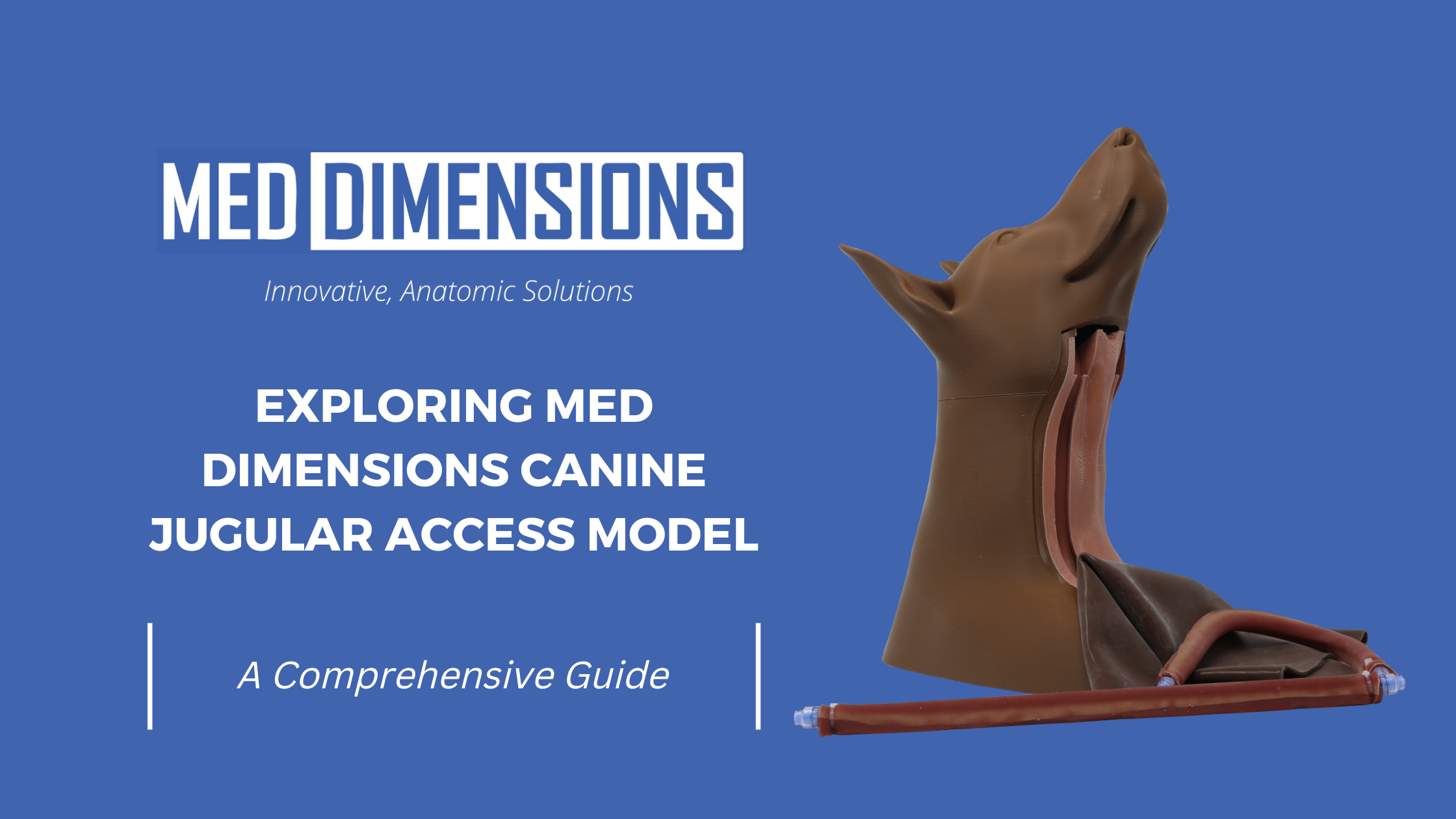
Understanding Med Dimensions Canine Jugular Access Model
Features:
- Realistic Anatomy: The Canine Jugular Access Model by Med Dimensions boasts lifelike canine anatomy, allowing practitioners to practice procedures in a highly accurate simulation.
- Durable Construction: Built with high-quality materials, the model is durable and designed to withstand repeated use, making it a cost-effective solution for training purposes.
- Longevity: During extensive training sessions, certain parts may need to be replaced which is why Med Dimensions offers replacement skins and vein sets at a discounted rate.
- User-Friendly: The model is user-friendly, providing a seamless experience for novice and experienced practitioners.
Performing Canine Jugular Access: A Step-by-Step Guide
Step 1: Preparation
Before beginning the procedure, gather all necessary equipment. This includes sterile gloves, an antiseptic solution, a jugular catheter, and a securing device.
Step 2: Restraint and Positioning
Secure the canine head in the correct recumbency. Proper positioning is crucial for successful jugular access. The head should be slightly elevated to distend the jugular vein.
Step 3: Aseptic Technique
Even though this is a training model, practicing strict aseptic techniques throughout the procedure is important for building good habits. This involves using sterile equipment and maintaining a clean environment to prevent infections.
Step 4: Locating the Jugular Vein
Palpate and visualize the jugular vein. It is typically found on the ventral aspect of the neck. Use gentle pressure to enhance visibility and make the vein more prominent.
Step 5: Insertion of the Catheter
Once the jugular vein is located, insert the catheter using a smooth, controlled motion. Be cautious to avoid complications such as hematoma or thrombosis.
Step 6: Securing the Catheter
Secure the catheter in place using an appropriate securing device. This ensures stability and reduces the risk of dislodgement.
Conclusion
The Med Dimensions Canine Jugular Access Model offers a valuable resource for veterinary professionals seeking to enhance their skills in a safe and controlled environment. By practicing jugular access, practitioners can ensure proficiency and confidence in this essential veterinary procedure. As technology continues to advance, such simulation models contribute significantly to the continuous improvement of veterinary care.