Anatomical Learning: The Crucial Role of Visual Aids and 3D Models
In the dynamic field of veterinary medicine, where precision and understanding of anatomical structures are paramount, the role of visual aids and 3D models cannot be overstated. Traditional methods of learning through textbooks and lectures are invaluable, but incorporating visual aids takes the educational experience to a whole new level. Med Dimensions is at the forefront of providing CT-derived models specifically designed for aiding the veterinary healthcare system.
- Enhanced Understanding and Retention:
Visual aids, such as 3D models, provide students with a three-dimensional perspective that surpasses the limitations of traditional two-dimensional illustrations. This immersive experience allows for a better understanding of the spatial relationships between organs, tissues, and structures within an animal’s body. Med Dimension’s Canine Arthroscopy Visual Stifle accurately replicates a true stifle and provides the elasticity in movement to replicate a living canine. This allows students to identify specific ligaments, their attachment points, and the range of motion that should be expected when maneuvering the hind legs of a canine. Research indicates that visual learning aids can significantly enhance retention and comprehension, making it an invaluable tool for veterinary students striving for excellence. - Interactive Learning Experience:
3D models enable students to interact with anatomical structures in a way that textbooks cannot replicate. The ability to rotate, zoom in, and manipulate the model provides a hands-on experience, fostering a deeper connection with the subject matter. This interactive learning experience engages students, making the study of veterinary anatomy more enjoyable and memorable. - Real-world Application:
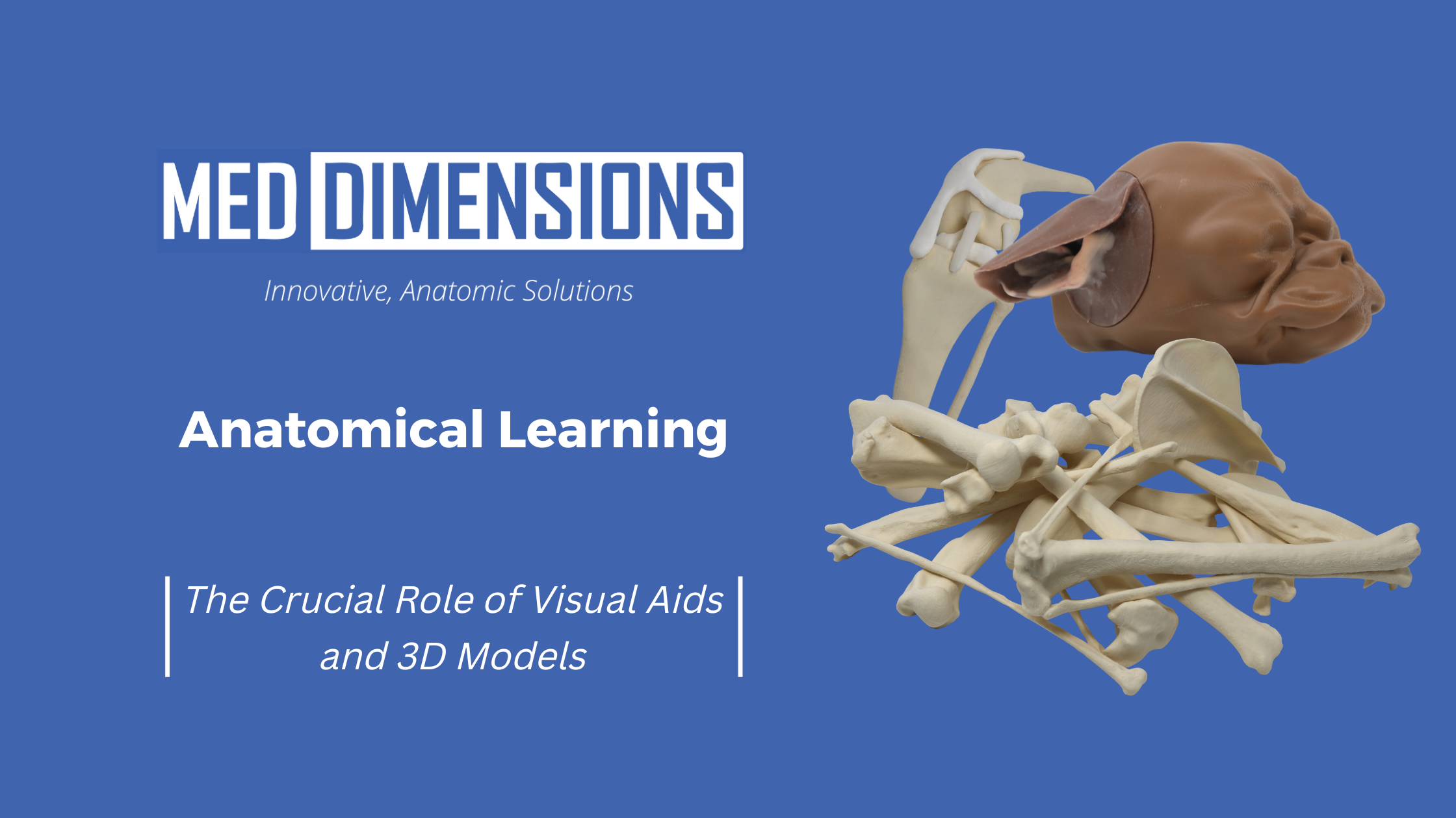
Veterinary medicine is a practical field, and the ability to apply theoretical knowledge to real-world scenarios is crucial. Visual aids, especially 3D models, bridge the gap between theory and practice. By simulating the complexities of an animal’s anatomy, students can develop a better grasp of how different structures function together in a living organism. The Med Dimensions Canine Ear model is ideal for using instruments such as the otoscope, where students can examine the ear canal before trying on a canine’s sensitive ear. - Effective Communication:
Veterinary professionals often collaborate with other members of the healthcare team, including clinicians, researchers, and even pet owners. The use of visual aids facilitates effective communication by providing a common visual reference point. Whether discussing surgical procedures, diagnostic findings, or treatment plans, the ability to convey complex anatomical information visually is an essential skill for veterinary practitioners. Med Dimensions also offers surgical models, 3D renderings and prints of deformed bones, or skeletons with the tumors included, providing a valuable resource for veterinary professionals to reference for surgery planning, and also as a communication method to share with pet owners on the severity and treatment plan options.
In the ever-evolving field of veterinary medicine, embracing visual aids and 3D models for anatomical learning is not just a luxury but a necessity. The benefits of enhanced understanding, interactive learning experiences, real-world application, effective communication, and the integration of technology underscore the importance of incorporating visual aids into veterinary education. By doing so, educational institutions empower the next generation of veterinary professionals with the knowledge and skills needed to excel in their careers and contribute to the well-being of animals.