VetBones: The Perfect Solution for Veterinary Procedures
Med Dimensions’ VetBones are changing the game for surgical practice.
We've Gone Global! International Shipping & Multiple Currencies Now Available Dismiss
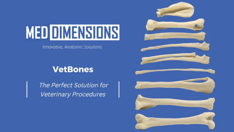
Med Dimensions’ VetBones are changing the game for surgical practice.
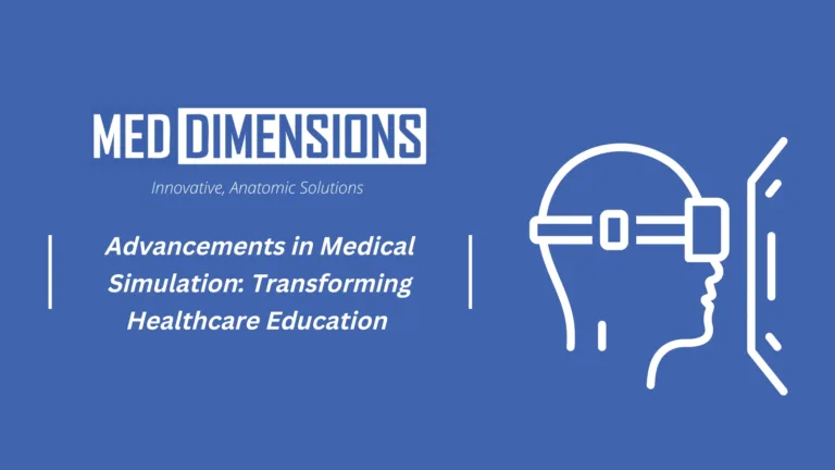
Introduction Over the past decade, medical simulation has emerged as a groundbreaking tool that has revolutionized healthcare education and training. By providing realistic and immersive experiences, medical simulation has significantly enhanced the skills and competence of healthcare professionals. From simulating complex surgeries to training for high-pressure emergency situations, medical simulation has proven to be a…
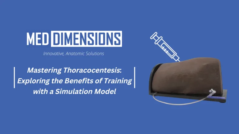
Thoracocentesis, a procedure involving the insertion of a needle into the pleural space to drain fluid or air, is a critical skill for veterinary professionals. It is vital for diagnosing and managing various respiratory conditions and emergencies. Proficiency in thoracocentesis requires knowledge, practice, and hands-on training. That’s where thoracocentesis training models come into play, providing…
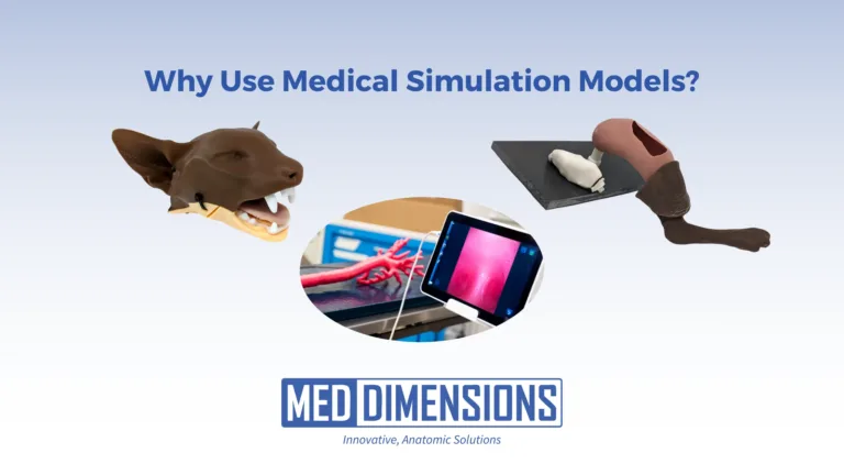
Medical simulation models are changing the way healthcare professionals are trained. They offer several advantages that improve patient outcomes, decrease errors, and enhance overall care. In this post, we will discuss the benefits of medical simulation models and how they are transforming healthcare education. Realistic Learning Environments Improved Clinical Outcomes Reduced Medical Errors Enhanced Teamwork…
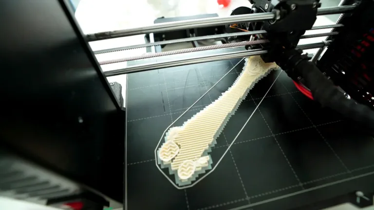
Do you ever wonder how companies can work with medical images in CAD programs? Here is a small peak behind the scenes how we at Med Dimensions are leveraging CAD and automation tools to create the next generation of Innovative Anatomic Solutions. Huge thanks to Gokce (Gilly) Yildirim from Vent Creativity for showing us the ropes. #meddimensions#innovation#caddesign
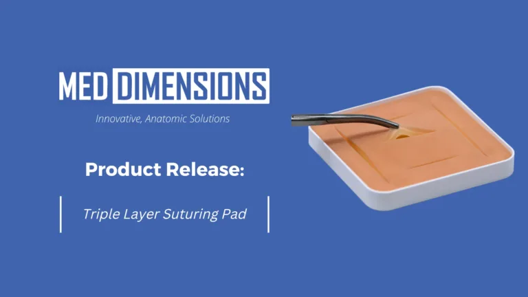
How do you place an ET when you can barely see the epiglottis? It’s not a rhetorical question; you practice! Med Dimensions is proud to offer 2 versions of high quality suturing pads for theteaching and learning of surgical suturing and knot tying: MD-E-0033C (3 slit) andMD-E-0053C (5 slit). Minimum ordering quantity is five (5)…
Annual Awards Program Recognizes Top Companies, Services and Products Within the Global Pet Industry
From Dr. Andrew Jackson Honey is a 1 year old female spayed Beagle mixed breed dog presented with a right forelimb angular limb deformity. Her deformity was quite pronounced compared to the left forelimb, which had mild typical valgus deformity. Radiographs revealed a biapical deformity of the right forelimb. I contacted Med Dimensions and presented…
An interview with Dr. Johnny Uday, a leading mind in innovative 3D medicine. 1) When did you know you wanted to get into veterinary medicine and helping animals? When I was a kid, we took our sick pet to the vet. I was so happy that my little dog was going to get help, and…
Med Dimensions produces 3D models for the education, preparation, and assistance of surgical procedures for veterinary doctors, clinicians, and teachers. From Chris Morgan at Matterhackers, Inc. – March 22, 2022 Located near Rochester, New York, Med Dimensions is a small startup created by two pet-loving engineers, Sean Bellefeuille and Will Byron, from Rochester Institute of…